Nachet Grand Modèle No. 1 - Perfectionné

Nachet Grand Modèle No. 1 or Grand Modèle Perfectionné
The Nachet Grand Modèle No. 1 (also termed the Grand Modèle Perfectionné) was widely regarded as the pinnacle of 19th-century French microscope design. While British makers favored sprawling, asymmetrical exhibition stands and German makers pushed for smaller, utilitarian laboratory benches, the Nachet Grand Modèle struck a balance as a prestigious, high-end flagship laboratory instrument.
The curated microscope is a rare late-19th/early 20th-century A. Nachet Grand modèle minéralogique polarizing microscope, Paris, circa 1886–1895, signed “A. Nachet, 17 rue Saint-Séverin, Paris,” brass and lacquered brass, approximately 51 cm high as configured, with inclined monocular body, heavy shaped horseshoe foot, twin support pillars, continental curved limb, rack-and-pinion coarse focus, fine focus adjustment, calibrated drawtube, large rotating graduated mineralogical stage, mechanical X–Y slide carrier, substage polarizing/condenser assembly, plane mirror, accessory objectives and eyepieces. Closely corresponding to the Nachet 1886 catalogue Grand modèle minéralogique.
This French Grand Modèle stands a highly impressive 20+ inches tall when deployed, fully extended for use.
The main microscope instrument, vertical illuminator, ultramicroscopic dark-field stage/condenser with spherical glasses/note, camera lucida, Nicol prisms/substage, cases, etc., overall condition is excellent for a 160+ year-old scientific instrument. This is a well-preserved, complete, and highly functional historical research outfit—rare in this degree of integrity.

A. NACHET
The A. NACHET Era: The business operated under several variations of its name. Camille Sébastien Nachet founded the company, followed by the partnership Nachet et Fils. However, when his son Alfred took sole control of the firm after Camille's death, he changed the engraving exclusively to A. NACHET. This specific naming convention was used roughly between 1880 and 1890.
The engraved address, 17, rue Saint-Séverin, Paris, was the location the workshop moved to in late 1861 and operated out of for decades.
By 1892, Alfred's own son Albert joined the business, and the signature permanently reverted back to Nachet et Fils.

Nachet Vertical Illuminator
The microscope is equipped with an uncommon internal prism lighting device signed "Nachet à Paris", in its original box signed "Nachet et fils, 17 rue St Sévrin Paris".
The Nachet Vertical Illuminator, also called Nachet prism vertical illuminator or Nachet incident-light / epi-illuminator for opaque objects is a specialized 19th-century accessory manufactured by Maison Nachet & Fils (A. Nachet et Fils), Paris, at their well-documented address 17 Rue St. Séverin. The device itself is signed “NACHET A PARIS”, and the original fitted case bears the full gold-stamped maker’s mark “NACHET & FILS / 17 RUE ST SEVERIN / PARIS” on the red velvet lining.
Key components and light path (matching the classic description in period sources such as Elementary Chemical Microscopy):
• Collimator tube (C): The long brass tube with a convex collecting lens (L) at the outer end. Light from an external source enters here.
• Prism cell (F): Contains a small right-angled reflecting prism (P) whose hypotenuse or relevant face (R) is silvered. The prism is positioned as close as possible to the back lens of the objective — a deliberate design feature that minimizes internal reflections, haze, and loss of contrast.
• Adapter (A): Threads into the lower end of the microscope body tube. The objective then screws directly into the illuminator’s lower threaded mount.
• Iris diaphragm: Adjustable via the side knob (K); can often be shifted eccentrically to optimize the point where light strikes the prism for different objectives/powers.
How it works:
1. External light (lamp, mirror-directed daylight, or modern LED/fiber-optic source) enters the collimator and is concentrated by the convex lens.
2. The silvered prism reflects the beam ~90° downward along the optical axis.
3. The objective now functions as a condenser, focusing a small, intense spot of light onto the specimen surface.
4. Light reflected or scattered from the opaque surface re-enters the objective and travels upward to form the image (most rays pass beside the small prism, which obstructs only part of the aperture).
5. Result: Dramatically brighter, higher-contrast views of surface details, textures, grain structure, inclusions, etch patterns, or bireflectance on gems/minerals compared with oblique or ambient lighting.
Advantages (period and practical):
• Excellent for moderate magnifications on opaque objects.
• Prism very close to objective → superior image quality with less flare/haze than many contemporary designs.
• Controllable intensity and effective aperture via iris.
• Transforms a transmitted-light polarizing microscope into a versatile instrument for reflected-light work.

Nachet Vertical Illuminator Above the Four Objective Nosepiece
The Grand Modèle Perfectionné No. 1 is equipped with a 4 Objective Nosepiece. The Nachet objective lens boldly up front in this view is one of 2 Imm Homog (Immersion Oil) high resolution lens. Note the excellent condition of all aspects of this microscope.
Centering Screws: If you look closely at the individual objective collar mounts on the nosepiece, you can see tiny adjustment screws. Petrographic work requires extreme precision; these allowed the user to micro-adjust each lens so that the center of the field of view never shifted when switching magnifications.

Nachet Camera Lucida
The specialized optical prism accessory mounted at the top of the microscope, signed “NACHET A PARIS”, was designed as a complementary fitting for Nachet compound or polarizing microscopes of the mid-to-late 19th century. It features a distinctive black-painted (or ebonized) rectangular/prism block with a circular aperture, an angled brass shield or deflector plate secured by screws, precision mounting threads/collar, and integrated optical elements (visible lens/prism face in the side view). The construction uses blackened components to minimize stray light, with brass fittings for durability and adjustment. This version of the camera lucida tilts and swivels, and also offers a deployable filter.
The Nachet top-mount camera lucida is a 19th-century optical drawing aid. It relies on a split-pupil or beam-splitting prism to superimpose a specimen’s magnified image onto a drawing surface. This allows a microscopist to trace the object exactly while keeping their eyes focused on the paper.
Key Design Features
Beam-Splitting Prism: Nachet’s design, famously improved by Dr. Govi around 1882, featured a larger prism with a thin layer of semi-transparent silver or gold. This allowed the observer to view the microscope's visual field and the sketching paper simultaneously with high clarity.
Top-Mount Configuration: The attachment clamps directly to the top of the microscope eyepiece. A laterally extending segment with a mirrored prism projects outward, casting the reflection of the drawing surface into the prism's beam splitter.
Split-Pupil Viewing: To use it effectively, the observer aligns their eye so that exactly half of the pupil is positioned over the prism and the other half observes the paper directly.
This accessory functions perfectly with Nachet’s polarizing microscope system such as this Large Model No. 1, where it supports advanced mineralogical, petrographic, or chemical observations by improving visibility of birefringent structures, inclusions, or textures under varied illumination alongside the vertical illuminator.

Nachet Accessory Components
The Grand Model No. 1 originally had a full lens array to choose from, including two oil immersion lens for high magnification.
The 1st & 3rd rack segments display Nachet’s standard dry objectives alongside his most expensive, high-numerical-aperture optical engineering marvels:
- The Homogeneous Immersion Lenses (\(1/15\) and \(1/18\)): The markings "imm homog" mean Immersion Homogène (Homogeneous Immersion). This technique was popularized by Ernst Abbe and Carl Zeiss around 1878–1880 to achieve extreme magnifications without optical distortion.
- Instead of leaving an air gap, a drop of specialized oil with the exact same refractive index as glass was placed between the slide cover slip and the tiny front glass of the lens.
- The fractions \(1/15\) and \(1/18\) represent the focal length of the lenses in Parisian inches (lignes). These were used for the absolute limits of 19th-century resolution—such as analyzing geological crystal structures, identifying blood cells, or diagnosing early bacterial pathogens like tuberculosis.
- Objectives 1, 3, 5, and 7: These are traditional low-to-high magnification dry objectives. The sequential numbering shows a flawlessly complete, original stepping progression from a low-power overview lens (No. 1) up to a high-power detail lens (No. 7).
The 2nd & 4th rack segments display Advanced Crystallography Optics
The bottom row reveals that this kit was intentionally optimized for advanced petrographic (rock/mineral) or polarizing analysis:
- Compens. Okular (Compensating Eyepieces): This German engraving format ("Compens. Okular" or Kompensations-Okular) refers to high-end compensating eyepieces. These were specially engineered with over-corrected color dispersion to perfectly counterbalance the chromatic aberrations naturally produced by high-power apochromatic and immersion objectives.
- Compensateur 1/4 (The Quartz Wedge/Wave Plate): The specialized brass tube labeled "Compensateur 1/4" is a quarter-wave plate (\(\lambda/4\) retarder). When dropped down into the optical path, it shifted the phase of light waves by exactly a quarter wavelength. Geologists used this to reveal subtle "interference colors" within mineral thin-sections, allowing them to instantly classify unknown crystal optical axes.

Nachet Grand Modèle No. 1 Vernier Mechanical Stage
The Grand Model No. 1 utilizes dual brass thumbscrews protruding from the circular stage, a large rotating graduated mineralogical stage. This is a precision Vernier mechanical stage, allowing micrometric horizontal and vertical movement of a glass slide. The stage also features a graduated degree scale along its rim, which indicates this model was likely configured for advanced petrographic (rock/mineral) and polarizing work, enabling full rotation of a specimen under examination.

Nachet Grand Modèle No. 1 Condenser Substage
The Substage Condenser Assembly: This is the complex cluster of brass underneath the stage. It includes an adjustable iris diaphragm and a condenser lens system, mounted on a rack-and-pinion track so you can precisely crank it up and down to focus light onto the slide.
The Dual-Sided Mirror: Located at the very bottom, the mirror is fully intact in near perfect condition. One side is flat (plane) and the other is concave to concentrate ambient light up through the condenser.

Nachet Grand Modèle No. 1 Substage Mirror
The Grand Model No. 1 utilizes a substage adjustable elevation swing-arm mirror, in near perfect condition, with an elevation knob on each side.
The gear mechanism is contained inside the box secured by four slotted bolts

Nachet Grand Modèle No. 1 Condenser Substage
The Grand Model No. 1 utilizes a substage fully adjustable condenser with Nicol prism. Below the substage plate is the condenser holder, mobile in the axis by means of a rack and can be eccentricated and separated laterally if you want to illuminate only with the mirror. The condenser receives at will the Abbe capacitor lighting that can be centered exactly. The iris diaphragm of the capacitor is mounted in rotation and can be moved laterally by means of a tangent screw, which makes it possible to obtain the oblique illumination by the capacitor.

Nachet Eclairage Ultramicroscopique Darkfield Accessory
The device exhibited is a specialized Nachet dark-field condenser / ultramicroscope stage accessory (labeled “Eclairage Ultramicroscopique” and “NACHET”), housed in its original fitted case with red velvet lining. It consists of a substantial brass annular mount with a central aperture, concentric rings (likely for light shaping/reflection), two silvered or adjustable levers/arms with knurled thumbscrews for precise centering and positioning, and a separate brass cap or cover (with black central element, possibly for protection, oil immersion contact, or specimen containment). The stage bears the serial number “120” and its signed “Nachet”.
Background and Historical Context
Nachet (a leading French microscope maker) produced this type of accessory in the early 20th century (post-1902/1903) to support ultramicroscopy, pioneered by Richard Zsigmondy and Henry Siedentopf. Traditional microscopes could not resolve particles smaller than ~0.2 μm (the diffraction limit). Ultramicroscopy uses intense dark-field illumination at very high oblique angles: light illuminates the specimen from the sides at angles too steep to enter the objective directly, creating a dark background. Sub-microscopic particles (e.g., colloidal gold, proteins, viruses, or emulsions) scatter light toward the objective, appearing as bright points or diffraction patterns (“stars on a black sky”) against darkness.
Nachet’s version was a practical, stage-mounted implementation for their compound microscopes, building on their expertise in condensers, prisms, and polarizing instruments. It complemented standard Abbe condensers and enabled chemical, colloidal, and biological research (e.g., Brownian motion studies by Perrin, Svedberg’s work).
Function - Produces extreme oblique (hollow-cone) illumination for dark-field ultramicroscopy:
- Core principle: Creates a hollow cone of highly oblique light (high numerical aperture, often via cardioid, paraboloid, or reflecting surfaces) focused on the specimen plane. Direct rays miss the objective; only scattered light from particles reaches the eyepiece.
- Dark-field effect: Background remains black; particles as small as a few nanometers become visible via Tyndall scattering.
- Ultramicroscopic capability: Extends resolution beyond ordinary limits for size/motion studies of colloids in suspension (not true imaging of shape, but detection and tracking).
- Light enters from below/sides and is shaped by internal reflecting surfaces into a very steep hollow cone.
- The cone’s apex focuses at the specimen plane. Direct rays miss the objective (creating a black background).
- Sub-microscopic particles scatter light into the objective, appearing as bright diffraction disks or points.
- High numerical aperture (often >1.2–1.4 with immersion) enables visualization of particles far below the normal resolution limit (~4–40 nm range for suitable specimens).
- The dual adjustment levers allow fine centering and optimization of the light cone for uniform illumination without glare.
Uses
- Colloid and nanoparticle research: Visualize and count gold sols, emulsions, proteins, or other sub-resolution particles in liquids.
- Live specimen observation: Blood, bacteria, or other suspensions without staining (preserves natural state).
- Chemical microscopy: Study precipitates, emulsions, or phase separations.
- Integration with Nachet polarizing microscopes: Enhances birefringence or scattering studies in transmitted light.
- Specimen prep: Typically a thin liquid film or drop between slide and coverslip (hence the note on “gouttes”/drops); often requires immersion oil between condenser and slide for highest obliquity.
Operation
- Mounting: Place the black base plate on the microscope stage (or in the substage holder). The central aperture aligns with the optical axis.
- Centering/Adjustment: Use the two knurled levers/screws to center the illuminating ring precisely and adjust height/focus. Fine tangent or eccentric controls optimize the light cone.
- Illumination Setup: Direct a strong light source (lamp, often with auxiliary condenser) into the device. Internal reflecting surfaces (concentric rings) form the oblique hollow cone.
- Immersion: Apply immersion oil or fluid to the top lens (or use the cap) for contact with the slide bottom to achieve maximum aperture and eliminate air interfaces.
- Observation: Use a low-to-medium NA objective (typically <1.0 to avoid capturing direct rays). Focus and observe scattered light points. Adjust for uniform dark background without glare.
- Cap/Cover: Protects optics or seals a specimen chamber/drop.
Performance Notes: Requires a powerful, well-collimated light source. Best with clean glassware and thin preparations. Modern equivalents use cardioid or paraboloid condensers; this Nachet piece remains a fine historical example of early ultramicroscopy hardware, valuable for collection/display or demonstration.
This accessory pairs well with other Nachet items (vertical illuminator, substage condenser, polarizing setup), expanding capabilities from standard brightfield/polarized to advanced dark-field/ultramicroscopic techniques. Condition appears excellent—original case and components intact.
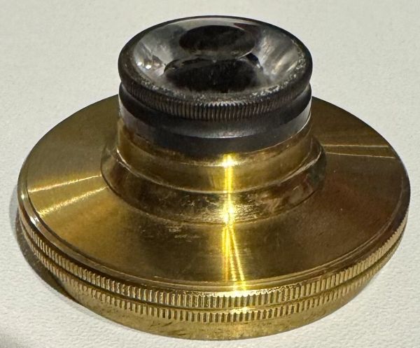
Nachet Ultramicroscopic Dark-Field Lens
This image exhibits the key optical element from the Nachet Eclairage Ultramicroscopique accessory: a specialized dark-field immersion lens/cap with a prominent central stop. This is the heart of the ultramicroscopic illumination system.
Technical Description
- Construction: A brass-mounted glass lens (likely plano-convex or specially figured) with a blackened/opaque central zone (the dark circular area in the center). The outer annular portion is clear, allowing light to pass only through the periphery. The rim is threaded or flanged for secure mounting onto the main condenser ring or as a top cap.
- Central stop: Deliberately opaque to block axial (straight-on) rays. This creates a hollow cone of highly oblique light—the defining feature of dark-field/ultramicroscopic illumination.
- Design purpose: When paired with the main brass condenser ring and adjustment plate, it ensures light strikes the specimen at steep angles (high numerical aperture, often requiring immersion oil contact). Direct rays are blocked, producing a black background; only scattered light from sub-microscopic particles reaches the objective.
Function in Operation
- Light path: Incoming light is shaped by the condenser’s concentric surfaces into a wide beam. The central stop eliminates the middle rays, forming an annular (ring-shaped) hollow cone that converges on the specimen plane at extreme obliquity.
- Immersion use: Oil (or appropriate fluid) is applied between this lens/cap and the underside of the slide for optical continuity and maximum aperture. This is why the note mentions “gouttes” (drops) alongside the matched optical glasses.
- Result: Sub-resolution particles (colloids, nanoparticles) scatter light sideways into the objective, appearing as bright points or halos. The field remains dark, dramatically increasing contrast.
This element works in tandem with the brass ring (concentric reflector) and black mounting plate depicted previously.
Background & Significance
Nachet adapted dark-field principles (building on earlier paraboloid/cardioid designs) for ultramicroscopy shortly after Zsigmondy and Siedentopf’s breakthrough (~1902–1903). This accessory allowed their polarizing and compound microscopes to visualize the invisible world of colloids, a major advance in physical chemistry and early nanotechnology. The complete set (condenser ring, adjustment plate, this lens/cap, glasses, case, and note) is a rare, intact example—most surviving pieces lack these precise optical elements.
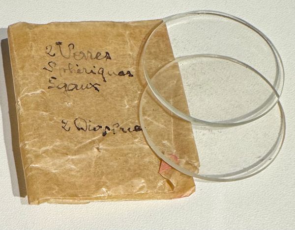
2 Verres Sphériques Egaux & 2 Dioptrie
• “2 Verres Sphériques [or Optiques] Egaux”: “2 equal spherical [or optical] glasses/lenses.”
• “2 Dioptrie”: Refers to two dioptric elements (lenses with specific optical power/focusing strength, measured in diopters).
The note is a quick French-language packing list or inventory slip for the spare matched pair of curved/spherical optical glasses (the two clear circular pieces you shared previously) plus their dioptric characteristics or associated elements. The paper shows age-related wear, creases, and a small pink/red seal or tape remnant, consistent with 19th/early 20th-century Nachet packaging.
Accessory Context
These are the matched spherical lenses used with the “gouttes” (drops) to create a thin, curved specimen chamber for ultramicroscopic observation. The curvature (spherical) improves light collection, immersion contact, and reduces aberrations in the high-oblique dark-field illumination—critical for visualizing sub-microscopic particles as bright scattering points (Tyndall effect) against a dark background.
Function and Role in Ultramicroscopy
These components enable proper specimen preparation for dark-field ultramicroscopy:
- Matched glasses: Sandwiched together with a small drop of liquid (the “gouttes”), they form a thin, uniform film or cell. This minimizes optical distortions and allows high-angle illumination without excessive scattering or absorption.
- Drops: The liquid medium suspends the sub-microscopic particles (colloids, nanoparticles, emulsions) for observation. Common in studies of Brownian motion or colloidal chemistry.
- Brass cap: Seals/protects the condenser optics or creates a stable contact point with the slide for oil immersion (essential for maximum numerical aperture and the steepest light cone).
Operation with These Items
- Place one optical glass on the stage over the condenser aperture.
- Add a small drop of specimen liquid (e.g., colloidal suspension) onto the glass.
- Cover with the second matched glass to form a thin, even layer (avoids air bubbles or uneven thickness).
- Use the brass cap/cover as needed for protection, oil contact, or additional focusing.
- Align and illuminate via the main condenser ring (as previously described). Particles appear as bright points against a dark background.
This setup is classic for early ultramicroscopy: clean, thin preparations are critical because even minor imperfections scatter light and ruin the dark field.

Dark-Field Lens / Cap (Central Stop Detail)
Nachet Ultramicroscopic Dark-Field Lens / Cap (Central Stop Detail)
This is the key optical element from the Nachet Eclairage Ultramicroscopique accessory: a specialized dark-field immersion lens/cap with a prominent central stop. This is the heart of the ultramicroscopic illumination system.
Technical Description
- Construction: A brass-mounted glass lens (likely plano-convex or specially figured) with a blackened/opaque central zone (the dark circular area in the center). The outer annular portion is clear, allowing light to pass only through the periphery. The rim is threaded or flanged for secure mounting onto the main condenser ring or as a top cap.
- Central stop: Deliberately opaque to block axial (straight-on) rays. This creates a hollow cone of highly oblique light—the defining feature of dark-field/ultramicroscopic illumination.
- Design purpose: When paired with the main brass condenser ring and adjustment plate, it ensures light strikes the specimen at steep angles (high numerical aperture, often requiring immersion oil contact). Direct rays are blocked, producing a black background; only scattered light from sub-microscopic particles reaches the objective.
Function in Operation
- Light path: Incoming light is shaped by the condenser’s concentric surfaces into a wide beam. The central stop eliminates the middle rays, forming an annular (ring-shaped) hollow cone that converges on the specimen plane at extreme obliquity.
- Immersion use: Oil (or appropriate fluid) is applied between this lens/cap and the underside of the slide for optical continuity and maximum aperture. This is why the note mentions “gouttes” (drops) alongside the matched optical glasses.
- Result: Sub-resolution particles (colloids, nanoparticles) scatter light sideways into the objective, appearing as bright points or halos. The field remains dark, dramatically increasing contrast.
This element works in tandem with the brass ring (concentric reflector) and black mounting plate you’ve shared previously. The two matched “Verres Optiques” (optical glasses) create the thin specimen chamber, and the drops suspend the particles.
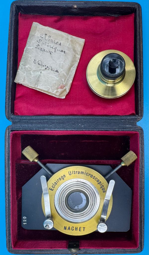
Nachet Eclairage Ultramicroscopique
The Nachet Eclairage Ultramicroscopique accessory: a specialized dark-field immersion lens with a prominent central stop. This is the heart of the ultramicroscopic illumination system.
Background & Significance
Nachet adapted dark-field principles (building on earlier paraboloid/cardioid designs) for ultramicroscopy shortly after Zsigmondy and Siedentopf’s breakthrough (~1902–1903). This accessory allowed their polarizing and compound microscopes to visualize the invisible world of colloids, a major advance in physical chemistry and early nanotechnology. The complete set (condenser ring, adjustment plate, this lens/cap, glasses, case, and note) is a rare, intact example—most surviving pieces lack these precise optical elements.
This rounds out an exceptional ultramicroscopy outfit that interoperates superbly with this Nachet polarizing microscope, vertical illuminator, Nicol prisms, and camera lucida.
This accessory group enables everything from mineral analysis to colloidal particle tracking—truly a comprehensive
historical research station.
Hayem–Nachet Hématimètre

Hayem–Nachet Hématimètre / Blood-Cell Counting Apparatus, Paris
Hayem–Nachet Hématimètre, Paris, late 19th century, c. 1875–1885.
Title: The Hayem–Nachet Hématimètre (Blood-Cell Counting Apparatus)
Maker: Maison Nachet (Signed Hayem & Nachet / A. Nachet)
Origin: 17, rue Saint-Séverin, Paris, France
Date: Circa 1880–1890 (A. Nachet Era)
Inventory Number: TBD
Description
An elite and remarkably complete late 19th-century clinical hematology kit housed in its original purple velvet- and silk-lined case, stamped “Hématimètre — Hayem & Nachet.” Co-designed by legendary French hematologist Dr. Georges Hayem and master builder Alfred Nachet, this apparatus represents the historic nineteenth-century pivot from purely descriptive blood microscopy to quantitative, diagnostic medicine. [1, 2]
The heart of this system is a specialized, Nachet-marked metal stage carrier housing a glass counting chamber with retaining clips. Rather than featuring standard, faint grid lines scratched into the slide surface—which historically vanished from view when submerged in liquid—this premium variant utilizes Nachet’s proprietary optical innovation: a dedicated, calibrated brass lens assembly containing a sharply visible photographic counting grid. The lens mechanism bolts directly underneath the slide platform, projecting the grid divisions directly into the optical path alongside the specimen to ensure flawless mathematical cell tracking.
A cased blood-cell counting apparatus associated with Professor Georges Hayem and Alfred Nachet, comprising a Nachet-marked metal cell carrier with glass counting chamber, retaining clips, brass optical lens assemblies, graduated glass pipettes, glass transfer tubes, cover glasses/lamellae, and fitted purple velvet-lined case stamped “Hématimètre — Hayem & Nachet.”
Designed for the numerical enumeration of blood corpuscles by diluting a measured blood sample, placing it in a calibrated chamber, viewing it microscopically against a defined grid or projected counting field, and converting the observed count into corpuscles per unit volume. A rare early medical-microscopy apparatus representing the transition from descriptive blood microscopy to quantitative hematology.

HÉMATIMÈTRE — HAYEM & NACHET
The Inventor: Georges Hayem (1841–1933) was a brilliant French physician who famously created "Hayem's Fluid" (mercuric chloride, sodium sulfate, and sodium chloride). This specific solution preserved red blood cells while preventing coagulation and clumping inside the counting apparatus.
The case is not incidental. It confirms the intended scientific function and gives the set a named medical-historical identity. The interior purple velvet and silk lining are consistent with Nachet’s high-grade accessory presentation

Lens with Engraved Grid
The major innovation was improving visibility of the counting grid. Nachet’s catalogue states that ordinary engraved divisions were hard to see when covered with liquid containing blood globules, and the Hayem–Nachet arrangement solved this by using a sharply visible photographic grid.

Lens with Engraved Grid
How the lens is used
The brass lens should be understood as part of the hematimeter counting system, not as a normal specimen-viewing objective for free use.
The likely use sequence is:
- Place the Nachet counting stage/cell on the microscope stage.
- Position the brass optical tube or lens system beneath the counting cell, aligned with the circular aperture.
- Illuminate from below using the microscope mirror or transmitted-light source.
- Bring the microscope objective into focus on the blood cells in the chamber.
- Adjust the hematimeter lens/grid system so the projected counting grid is visible in the same field as the blood corpuscles.
- Count the cells within the defined grid divisions.
- Rotate or adjust only as necessary; the key is stable alignment between the projected grid, the chamber, and the microscope optical axis.
The close-up showing the square aperture inside the brass lens strongly suggests that this component contains or images the counting-grid element. When viewed through the microscope, that grid would establish the field divisions for counting. The operator would not normally hold this lens by hand like a loupe. It is a calibrated optical component meant to work in fixed relationship to the cell.

Lens with Engraved Grid
How the lens is used
The brass lens should be understood as part of the hematimeter counting system, not as a normal specimen-viewing objective for free use.
The likely use sequence is:
- Place the Nachet counting stage/cell on the microscope stage.
- Position the brass optical tube or lens system beneath the counting cell, aligned with the circular aperture.
- Illuminate from below using the microscope mirror or transmitted-light source.
- Bring the microscope objective into focus on the blood cells in the chamber.
- Adjust the hematimeter lens/grid system so the projected counting grid is visible in the same field as the blood corpuscles.
- Count the cells within the defined grid divisions.
- Rotate or adjust only as necessary; the key is stable alignment between the projected grid, the chamber, and the microscope optical axis.
The close-up showing the square aperture inside the brass lens strongly suggests that this component contains or images the counting-grid element. When viewed through the microscope, that grid would establish the field divisions for counting. The operator would not normally hold this lens by hand like a loupe. It is a calibrated optical component meant to work in fixed relationship to the cell.
Lens Mounted
Specimen slide is positioned over the lens.

Lens with Engraved Grid
The slide platform is held onto the microscope stage by its clips. The Hematimetre lens bolts onto the bottom of its stage.

Lens with Engraved Grid
The Hematimetre lens bolts onto the bottom of its stage as seen.

Lens with Engraved Grid
The slide platform is held onto the microscope stage by its clips. The Hematimetre lens bolts onto the bottom of its stage.

Lens with Engraved Grid
This view of the underside of the Hématimètre stage exhibits the special grid engraved insert through which is illuminated from the microscope base mirror reflected light is cast up through the specimen blood sample, thus enabling a precision count of blood cells including Red Blood Cells (Erythrocytes)
& White Blood Cells (Neutrophils, Lymphocytes, Monocytes, Eosinophils, Basophils). Differentiation of white blood cell types was not likely possible until later.